Spread of misfolded proteins could trigger type 2 diabetes

Type 2 diabetes and prion disease seem like an odd couple, but they have something in common: clumps of misfolded, damaging proteins.

Now new research finds that a dose of corrupted pancreas proteins induces normal ones to misfold and clump. This raises the possibility that, like prion disease, type 2 diabetes could be triggered by these deformed proteins spreading between cells or even individuals, the researchers say.

When the deformed pancreas proteins were injected into mice without type 2 diabetes, the animals developed symptoms of the disease, including overly high blood sugar levels, the researchers report online August 1 in the Journal of Experimental Medicine.
“It is interesting, albeit not super-surprising” that the deformed proteins could jump-start the process in other mice, says Bruce Verchere, a diabetes researcher at the University of British Columbia in Vancouver. But “before you could say anything about transmissibility of type 2 diabetes, there’s a lot more that needs to be done.”

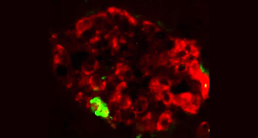
Beta cells in the pancreas make the glucose-regulating hormone insulin. The cells also produce a hormone called islet amyloid polypeptide, or IAPP. This protein can clump together and damage cells, although how it first goes bad is not clear. The vast majority of people with type 2 diabetes accumulate deposits of misfolded IAPP in the pancreas, and the clumps are implicated in the death of beta cells.

Deposits of misfolded proteins are a hallmark of such neurodegenerative diseases as Alzheimer’s and Parkinson’s as well as prion disorders like Creutzfeldt-Jakob disease (SN: 10/17/15, p. 12).

Since IAPP misfolds like a prion protein, neurologist Claudio Soto of the University of Texas Health Science Center at Houston and his colleagues wondered if type 2 diabetes could be transmitted between cells, or even between individuals. With this paper, his group “just wanted to put on the table” this possibility.

The mouse version of the IAPP protein cannot clump — and mice don’t develop type 2 diabetes, a sign that the accumulation of IAPP is important in the development of the disease, says Soto. To study the disease in mice, the animals need to be engineered to produce a human version of IAPP. When pancreas cells containing clumps of misfolded IAPP, taken from an engineered diabetic mouse, were mixed in a dish of healthy human pancreas cells, it triggered the clumping of IAPP in the human cells.
The same was true when non-diabetic mice got a shot made with the diabetic mouse pancreas cells. The non-diabetic mice developed deposits of clumped IAPP that grew over time, and the majority of beta cells died. When the mice were alive, more than 70 percent of the animals had blood sugar levels beyond the healthy range.

Soto’s group plans to study if IAPP could be transmitted in a real world scenario, such as through a blood transfusion. They’ve already begun work on transfusing blood from mice with diabetes to healthy mice, to see if they can induce the disease. “More work needs to be done to see if this ever operates in real life,” Soto says.

Even if transmission of the misfolded protein occurs only within an individual, “this opens up a lot of opportunities for intervention,” Soto says, “because now you can target the IAPP.”

Verchere also believes IAPP is “a big player” in the progression of type 2 diabetes, and that therapies that prevent the clumps of proteins from forming are needed. Whether or not future research supports the idea that the disease is transmissible, the study is “good for appreciating the potential role of IAPP in diabetes.”

Normally aloof particles of light seen ricocheting off each other

Cross two flashlight beams and they pass right through one another. That’s because particles of light, or photons, are mostly antisocial — they don’t interact with each other. But now scientists have spotted evidence of photons bouncing off other photons at the Large Hadron Collider at CERN, the European particle physics lab in Geneva.

“This is a very basic process. It’s never been observed before, and here it is finally emerging from the data,” says theoretical physicist John Ellis of King’s College London who was not involved with the study. Researchers with the ATLAS experiment at the LHC report the result August 14 in Nature Physics.
Because photons have no electric charge, they shouldn’t notice one another’s presence. But there’s an exception to that rule. According to quantum mechanics, photons can briefly transform into transient pairs of electrically charged particles and antiparticles — such as an electron and a positron — before reverting back to photons. Predictions made more than 80 years ago suggest that this phenomenon allows photons to interact and ricochet away from one another.

This light-by-light scattering is extremely rare, making it difficult to measure. But photons with more energy interact more often, providing additional chances to spot the scattering. To produce such energetic photons, scientists slammed beams of lead nuclei together in the LHC. Photons flit in and out of existence in the lead nuclei’s strong electromagnetic fields. When two nuclei got close enough that their electromagnetic fields overlapped, two photons could interact with one another and be scattered away.

To measure the interaction, ATLAS scientists sifted through their data to find collisions in which only two photons — the two that scattered away from the collision — appeared in the aftermath. “That’s the trickiest part of the whole thing,” says physicist Peter Steinberg of Brookhaven National Laboratory in Upton, N.Y., a member of the ATLAS collaboration. The scientists had to ensure that, in their enormous, highly sensitive particle detector, only two photons appeared, and convince themselves that no other particles had gone unaccounted for. The researchers found 13 such events over 19 days of data collection. Although other processes can mimic light-by-light scattering, the researchers predict that only a few such events were included in the sample.

The number of scattering events the researchers found agrees with the predictions of the standard model, physicists’ theory of particle physics. But a more precise measurement of the interaction might differ from expectations. If it does, that could hint at the existence of new, undiscovered particles.

These chip-sized spacecraft are the smallest space probes yet

Spacecraft have gone bite-sized. On June 23, Breakthrough Starshot, an initiative to send spacecraft to another star system, launched half a dozen probes called Sprites to test how their electronics fare in outer space. Each Sprite, built on a single circuit board, is a prototype of the tiny spacecraft that Starshot scientists intend to send to Alpha Centauri, the trio of stars closest to the sun. Those far-flung probes would be the smallest working spacecraft yet.

“We’re talking about launching things that are a thousand times lighter than any previous spacecraft,” says Avi Loeb, an astrophysicist at Harvard University who is part of the committee advising the initiative. A Sprite is only 3.5 centimeters square and weighs four grams, but packs a solar panel, radio, thermometer, magnetometer for compass capabilities and gyroscope for sensing rotation.

These spacecraft are designed to fly solo, but for this test, they hitched a ride into low Earth orbit on satellites named Max Valier and Venta-1. Each satellite has one Sprite permanently riding sidecar, and the Max Valier craft has another four it could fling out into space. Unfortunately, as of August 10, ground controllers haven’t yet been able to reach the Max Valier satellite to send a “Release the Sprites!” command. One of the permanently attached Sprites — probably the one on Venta-1 — is in radio contact.

Before sending next-gen Sprites off to Alpha Centauri, scientists plan to equip them with cameras, actuators for steering and other tools. “This was really just the first step in a long journey for Starshot,” Loeb says.

This sea snake looks like a banana and hunts like a Slinky

With its bright hue, this snake was bound to stand out sooner or later.

A newly discovered subspecies of sea snake, Hydrophis platurus xanthos, has a narrow geographic range and an unusual hunting trick. The canary-yellow reptile hunts at night in Golfo Dulce off Costa Rica’s Pacific coast. With its body coiled up at the sea surface, the snake points its head under the water, mouth open. That folded posture “creates a buoy” that stabilizes the snake so it can nab prey in choppy water, says study coauthor Brooke Bessesen, a conservation biologist at Osa Conservation, a biodiversity-focused nonprofit in Washington, D.C. In contrast, typical Hydrophis platurus, with a black back and yellow underbelly, hunts during the day, floating straight on calm seas.
The newly described venomous snake has been reported only in a small, 320-square-kilometer area of Golfo Dulce. After analyzing 154 living and preserved specimens, the researchers described the reptile’s characteristics July 24 in Zookeys. The scientists hope that the subspecies designation will enable the Costa Rican government to protect the sunny serpent, which they worry is already at risk from overzealous animal collectors.

Coconut crabs are a bird’s worst nightmare

Imagine you’re a red-footed booby napping on a not-quite-high-enough branch of a tree. It’s nighttime on an island in the middle of the Indian Ocean, and you can’t see much of what’s around you. Then, out of the darkness comes a monster. Its claw grabs you, breaking bones and dragging you to the ground. You don’t realize it yet, but you’re doomed. The creature breaks more of your bones. You struggle, but it’s a fruitless effort. Soon the other monsters smell your blood and converge on your body, ripping it apart over the next few hours.

The monster in this horror-film scenario is a coconut crab, the world’s largest terrestrial invertebrate, which has a leg span wider than a meter and can weigh more than four kilograms.

But this is no page from a screenplay. Biologist Mark Laidre of Dartmouth University actually witnessed this scene in March 2016, during a two-month field expedition to study the crabs in the Chagos Archipelago.

Laidre, an expert on hermit crabs, had been “dying to study” their humongous cousins. Little is known about the crabs, he notes. A study earlier this year looked at the force a coconut crab’s claw can exert in the lab. But, he says, “there’s still not a single paper on how they open a coconut.”
He trekked to the remote spot in the Indian Ocean because he wanted to study the crabs in a place where few people would interfere with their natural behaviors. Laidre had heard stories that coconut crabs killed rats, and he later witnessed them munching on the rodents on the islands. “Clearly it’s in their repertoire to eat something big,” he says. And when he took inventory of the crabs’ burrows, he found the carcass of an almost full-grown red-footed booby in one. “At the time, I had assumed it was something that had died … and the crab had dragged in there,” he recalls.

But then, in the middle of the night, he saw a crab attack a bird sleeping in a tree, and he managed to catch part of the event on film. “I didn’t have the heart to videotape five coconut crabs tearing apart the bird later,” he says. “It was a little bit overwhelming. I had trouble sleeping that night.”
After the event, Laidre heard a story from a local plantation worker who had witnessed something similar a couple of years earlier. “He was sitting and eating a sandwich, and this coconut crab came right out its burrow in the middle of the daytime when … a red-footed booby… landed outside of its burrow,” Laidre says. The crab grabbed the bird’s leg and pulled it into the burrow. “The bird never emerged.”

It’s difficult to tell how often attacks like this happen, whether they’re rare or common. “Predation itself is something you don’t often witness,” Laidre says. He’d like to someday install camera traps on the islands to get a better sense of the crabs’ behavior.

But while he was in the Chagos, he did find himself in a sort of natural experiment that gave him some insight into the effect of the crabs on local bird populations. Coconut crabs live on only some of the islands. Birds can live on any of them, but their populations vary from island to island. So Laidre surveyed the islands, walking transects and counting crabs and bird nests.
“The pattern I found across the island was pronounced,” Laidre writes November 1 in Frontiers in Ecology and the Environment. On Diego Garcia, for example, a 15-kilometer transect revealed 1,000 crabs and no nesting birds. Crab-free West Island, in contrast, had an abundance of ground nests of nesting noddies.

Laidre suspects that the coconut crabs act as a “ruler of the atoll,” keeping ground-nesting bird species from finding homes on crab-filled islands. On other islands with large populations of birds, those birds might help to keep their islands crab-free by eating juvenile coconut crabs, preventing them from colonizing there.

“It’s easy to sympathize with the prey,” Laidre says, “but at the same time, there’s a lot of ecological roles that that sort of action has.”

These disease-fighting bacteria produce echoes detectable by ultrasound

Ultrasound can now track bacteria in the body like sonar detects submarines.

For the first time, researchers have genetically modified microbes to form gas-filled pouches that scatter sound waves to produce ultrasound signals. When these bacteria are placed inside an animal, an ultrasound detector can pick up those signals and reveal the microbes’ location, much like sonar waves bouncing off ships at sea, explains study coauthor Mikhail Shapiro, a chemical engineer at Caltech.

This technique, described in the Jan. 4 Nature, could help researchers more closely monitor microbes used to seek and destroy tumors or treat gut diseases (SN: 11/1/14, p. 18).
Repurposing ultrasound, a common tissue-imaging method, to map microbes creates “a tool that nobody thought was even conceivable,” says Olivier Couture, a medical biophysicist at the French National Center for Scientific Research in Paris, who wasn’t involved in the work.

Until now, researchers have tracked disease-fighting bacteria in the body by genetically engineering them to glow green in ultraviolet images. But that light provides only blurry views of microbes in deeper tissue — if it can be seen at all. With ultrasound, “we can go centimeters deep and still see things with a spatial precision on the order of a hundred micrometers,” Shapiro says.

Story continues below image
Shapiro and his colleagues engineered a strain of E. coli used to treat gut infection to form gas compartments, and injected these bacteria into mice’s bellies. Unlike glowing bacteria — which could only be pinpointed to somewhere in a mouse’s abdomen — ultrasound images located the gas-filled microbes in the colon. The researchers also used their ultrasound technique in mice to image Salmonella bacteria, which could be used to deliver cancer-killing drugs to tumor cells.

Bacteria that produce ultrasound signals can also be designed to help diagnose illnesses, Shapiro says. For instance, a patient could swallow bacteria engineered to create gas pockets wherever the microbes sense inflammation. A doctor could then use ultrasound to search for inflamed tissue, rather than performing a more invasive procedure like a colonoscopy.