Reef rehab could help threatened corals make a comeback

Coral reefs are bustling cities beneath tropical, sunlit waves. Thousands of colorful creatures click, dash and dart, as loud and fast-paced as citizens of any metropolis.

Built up in tissue-thin layers over millennia, corals are the high-rise apartments of underwater Gotham. Calcium carbonate skeletons represent generations of tiny invertebrate animals, covered in a living layer of colorful coral polyps. Their structures offer shelter, and for about 114 species of fish and 51 species of invertebrates, those coral skyscrapers are lunch.

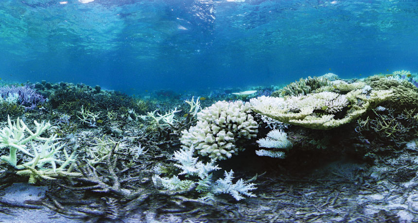
Important as they are, corals are in jeopardy. Warming oceans are causing more and more corals to bleach white and become vulnerable to destruction. A prolonged spike in temperatures, just 1 to 2 degrees Celsius, is enough to kill the marine animals. Greenhouse gas emissions also acidify the water, dissolving the calcium skeletons. In some countries, fishermen use dynamite to catch fish, leaving behind coral rubble. Today, more than 60 percent of the world’s reefs are at risk of disappearing.
Threats to reefs have “dramatically escalated in the last few decades,” says marine scientist Peter Harrison of Southern Cross University in Lismore, Australia. He has studied corals for three decades. “In my time as a reef researcher,” Harrison says, “I’ve seen it get worse, firsthand.”

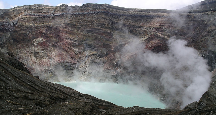
Thirty years ago, massive coral bleachings were unheard of. Today, reefs are suffering through a third global bleaching event since 1998. With high ocean temperatures dragging on since 2014, this summer marked the longest and most widespread episode of worldwide coral bleaching on record (SN: 7/23/16, p. 5). Australia has been hit especially hard. More than 80 percent of the northern part of the Great Barrier Reef is bleached and close to half of those corals have died, according to a report in April from Australia’s National Coral Bleaching Taskforce.

As reefs take a nose dive, scientists from Hawaii to the Philippines and the Caribbean are scrambling to save corals. Approaches that were once considered radical are “now seen as necessary in some places,” says coral biologist Ruth Gates of the Hawaii Institute of Marine Biology on Oahu.

In Florida, researchers are restoring reefs with tiny coral fragments. In Hawaii, Gates is scouring the water for stress-tolerant corals and experimenting in the lab to breed the hardiest individuals. At the 13th International Coral Reef Symposium in Honolulu in June, Harrison’s team reported early promising results of its effort to flood damaged reefs in the Philippines with tiny coral larvae.

What works on one reef won’t necessarily save another. So researchers are testing an arsenal of options to rescue a diversity of underwater communities.

A different story
In the early 1980s, Harrison was a graduate student at James Cook University in Townsville, Australia, working on the Great Barrier Reef. At the time, textbooks taught that most corals reproduce by brooding: Fertilization occurred inside the body and larvae were released into the water to replenish reefs year-round. But Harrison witnessed something very different. For a few nights around a full moon in springtime, corals spawned, spewing eggs and sperm into the water to be externally fertilized. The sea was covered in a pink, oily slick.

“We found the corals hadn’t read the textbooks,” Harrison says. Eggs and sperm were meeting outside of the coral bodies, and larvae were developing while drifting in the currents.

That discovery spurred a cascade of studies on coral reproduction that led to the modern understanding that many corals reproduce only once or twice a year, in coordinated mass releases of eggs and sperm. Most of the resulting larvae die or drift out to sea, Harrison says. Only a small fraction survive to adulthood. Even so, mass spawns are “how reefs replenish themselves over time,” he says.

Just after Harrison’s discovery, the Great Barrier Reef, and then reefs around the globe, experienced bleaching on a massive scale. Normally, tiny algae live inside coral polyps. The algae make sugar and other nutrients for the coral, and can give polyps their characteristic bright colors. But when temperatures spike, algae become toxic. Corals spit out their partners, bleach white and can die if temperatures don’t cool enough for the algae to return (SN Online: 10/8/15).

Corals worldwide were bleaching more often and more severely than had been recorded in the past. Scientists began to worry that reefs were in trouble. Some researchers, like Dave Vaughan, who manages the Coral Reef Restoration program at the Mote Tropical Research Laboratory in Summerland Key, Fla., took action.

In those days, Vaughan was a fish farmer, raising saltwater fish species in captivity. He began growing corals for tropical aquarium tanks. At the time, all the corals in the aquarium trade were taken from the wild, Vaughan says. He started growing coral species in captivity as an environmentally friendly alternative.

One day, Philippe Cousteau, grandson of legendary aquanaut Jacques, toured the operation. When the young Cousteau saw that Vaughan was raising corals for aquariums, “he shook his head,” Vaughan remembers, “and said ‘Dave, if you could do this for the aquarium trade, you can do this for the reef.’”

In those earliest days, most scientists were tackling small-scale reef damage caused by dropped anchors or boat groundings, Vaughan says. To repair that kind of minor damage, scientists began breaking 3- to 5-centimeter fragments from healthy corals on a neighboring reef and transplanting the chunks in damaged spots.

Cousteau’s visit convinced Vaughan that he should try restoring reefs. While doing so, 11 years ago, Vaughan made a game-changing discovery: Tinier fragments of coral, only 1 centimeter long, repair themselves 25 to 40 times as fast as scientists had ever recorded corals growing.

Today Vaughan’s team is spreading many of these microfragments over the surface of dead coral skeletons in the Florida Keys. As those bits fuse back together, they create a fast-growing “skin” over an otherwise dead reef. Condemned buildings are refurbished rather than razed.

The hope is that thousands of microfragments will carpet a small reef in two to three years, says Chris Page, a biologist at Mote Marine Laboratory working with Vaughan. That’s super fast. “There’s no way that’s happening in nature,” Page says.

Vaughan’s team is cultivating 17 species for microfragmentation in large troughs on land, with seawater running through them. He is focused on the top six slow-growing massive species that create the foundation of the reef. Some can live for centuries, mounded into boulders the size of a truck.
The Florida researchers plunged their first 200 microfragments into the ocean three years ago, at two sites in a nearshore coral reef off Big Pine Key, Fla. The colonies are now six to eight times as large as they were at planting and have begun to fuse together into areas about the size of a 5-gallon bucket lid. Since then, Vaughan and Page have planted close to 10,000 microfragments in the wild. “People were looking for some glimmer of light,” Vaughan says. “And restoration is turning out to be that in a big way.”

Seeds of reefs
Fragmentation and the newer microfragmentation are both time- and labor-intensive, and therefore very expensive. And, Harrison says, they rely on cloning.

When one coral is broken into fragments, to be fattened up and then planted around a reef, each chunk is genetically identical. All those pieces have the same DNA blueprint to fight infection and to deal with stress. Unlike natural reefs, where individuals are genetically distinct and have different vulnerabilities, cloned corals share the same weaknesses.

“People have spent years growing coral gardens only to have them wiped out by the next bleaching event,” Harrison says. With more diversity, he adds, some of those corals might have survived. In a warmer world where bleaching and disease will probably become more common, “genetic diversity equals resilience.”

To address the diversity issue, Vaughan and Page are raising 20 to 30 genetic variants of each coral species, to be planted around the reef. They are also collecting eggs and sperm from wild colonies of four coral species to grow on Summerland Key.

Harrison has been thinking about genetic diversity ever since the early 1980s, when he saw corals spewing sperm and eggs into the ocean. Few of the resulting larvae would survive. Many would drift away and most would die. All while Harrison saw reefs in decline.

What if, he wondered, scientists could take millions of those diverse coral larvae and help them settle onto reefs to replenish ailing ecosystems?

Other researchers asked themselves the same question. In the late 1990s and from 2007 to 2009, two teams, in Australia and Palau, released coral larvae onto healthy reef areas in mesh tents pitched over the seabed. In both studies, thousands of larvae settled under the tents, many more than scientists would have seen naturally.

But those early results may have been misleading. Most of the early settlers in Palau died within 30 weeks. Flooding the reef with larvae didn’t make a lasting difference in coral numbers. Maybe, the researchers speculated, settlers were too crowded, which meant swamping reefs with larvae made no sense.

Harrison wasn’t ready to give up. Even though most of the new settlers had died, those studies were done on healthy reefs, he says. In battered areas, where some baby corals might naturally drift in, but not enough for the reef to self-heal, a flush of larvae could be a shot in the arm.

The idea was to find a badly damaged reef, where the worst problems, such as blast fishing, had stopped. Harrison would bring a few of the reef’s mature, sexually active corals to the lab, persuade them to release sperm and eggs in aquarium tanks, and then take more than a million of their larvae back out to the reef. The plan was to saturate the environment with settling babies, as adult corals would have done in healthier days.

In 2013, Harrison’s team, led by graduate student Dexter dela Cruz, began a small pilot experiment in the Philippines at a reef called Magsaysay, where nearly two decades of fishing with explosives had taken a toll. Blast fishing is “like hitting the reef with a sledgehammer,” Harrison says. Magsaysay’s large foundational corals were blown to bits. A once-vibrant city was now a wasteland.
By 2013, the blast fishing had stopped, but Magsaysay wasn’t recovering on its own. So Harrison’s team brought in larvae from a species of fast-growing, purple-tipped coral, called Acropora tenuis, collected from a nearby healthier reef. The scientists released more than a million larvae into floorless mesh tents pitched under-water over the reef. After five days, Harrison’s team removed the mesh enclosures.

Over the next six months, most of the tiny coral settlers died. But, by the nine-month mark, the remaining populations had stabilized. Scientists expected more of the juvenile corals to die, but “incredibly and extraordinarily,” Harrison says, none have. At 3 years old, the juvenile corals have reached sexual maturity and are now the size of dinner plates. In June, dela Cruz presented the findings in Honolulu.

For slower-growing corals, Harrison’s approach will take extra patience. But for fast-growers like A. tenuis, reseeding larvae could be a quick and affordable way to help severely damaged reefs bounce back.

Winning corals
Getting more larvae onto damaged reefs is the first step, Harrison says. But some individuals are stronger and more stress-tolerant than others. As they grow up, these “winners” distinguish themselves by surviving.

Across the Pacific from Magsaysay, biologist Gates is studying winners. Rows of indoor and outdoor aquariums gurgle in her lab on Coconut Island, off Oahu’s windward shore. Those tanks are full of Montipora capitata, a local and fast-growing coral collected from the patchy reefs surrounding the island.

In 2014 and 2015, unusually warm water hit Hawaii. Under stress, many corals rejected their symbiotic algae, then blanched from a healthy brown to white; some died.

Gates’ team patrolled the reefs around the island during and after the bleaching, in search of hardy M. capitata individuals that stayed brown, even in hot water. The scientists are also interested in M. capitata that bleached, but then recovered. Gates equates the work to professional sports scouting, “out at high schools, looking for the best athletes.”
When she finds top performers, Gates brings them to her lab to run them through their paces, exposing each pro-performer to different temperatures and pH levels in seawater tanks. Some conditions re-create today’s oceans, while others mimic future warm and more acidic seas.

Today, Gates is breeding the strongest corals (her first batch of babies was born in June). She hopes that top performers will have “extremely talented kids” that inherit their parents’ strengths. It’s too soon to tell how the new corals will do once they’re planted out on the reef.

“We’re trying to give corals a leg up,” she says. Reefs healthy enough to survive without human intervention are the ultimate aim. In the next five years, the researchers plan to branch out from M. capitata to look for super corals of all five species found in the bay surrounding Coconut Island.

It would be ideal to find those super corals before the next big bleaching event. But for that, the researchers need another sign of resilience. That sign, Gates says, could be hidden in the way corals glow.

Some coral animals, and their symbiotic algae, are loaded with fluorescent proteins that absorb incoming light, then spit it back out by glowing. It’s unclear what fluorescent proteins do for corals; they may be a kind of sun block, protecting corals from the intense light in shallow seas, or a form of camouflage or part of the immune system.

Stress affects corals’ glowing proteins and changes their fluorescence patterns. In the Pacific and Indian Ocean species Acropora yongei, for instance, researchers reported in 2013 in Scientific Reports that the concentration of green fluorescent protein fell with temperature stress before bleaching and the coral glowed less intensely. In an earlier study, prolonged high temperatures changed the ratio of green to orange fluorescence in the endangered Caribbean coral Orbicella faveolata.

Gates expects that under stress, super corals will keep their healthy fluorescence patterns much longer than corals that are bleaching. One next step, Gates says, is to stress out tiny pieces of coral and watch what happens under a very powerful laser scanning confocal microscope. She’ll expose nubbins of coral to acidifying water or increasing temperatures in a petri dish. The microscope will pick up the fluorescence of the nubbins and may indicate which corals will stay healthy the longest.
Once scientists can identify the hardiest corals, they can combine selective breeding with other rehab techniques. Approaches like microfragmentation could help super colonies mature super fast. Then, Gates says, “we would have a strategy to get the reef producing its own offspring quite quickly.”

No two approaches to saving reefs are the same, which is probably a good thing. Coral fragmentation, reseeding and selective breeding each have their pros and cons.

“The assumption that one size will fit all is completely flawed,” Gates says. What might work on the Florida coast wouldn’t necessarily work in the Pacific. Like far-flung cities, each reef has different needs and priorities. Their communities of coral vary as do the threats they face. Some problems, like warming oceans, are global in scope. Others, like pollution from roads and agricultural runoff, overfishing and dynamite fishing, are often more localized. Rehabilitation approaches will vary, depending on the type and severity of damage, and how the mosaic of coral species might respond.

Across the globe, “will the things that we do be different?” Gates asks. “Absolutely.”

Rather than competing, Gates, Vaughan and Harrison are working toward a common goal: to find the right mix of approaches to support the reefs so they no longer need human help.

There’s a new way to stop an earthquake: put a volcano in its path

Editor’s note: Science has retracted the study described in this article. The May 3, 2019, issue of the journal notes that a panel of outside experts convened by Kyoto University in Japan concluded in March 2019 that the paper contained falsified data, manipulated images and instances of plagiarism, and that these were the responsibility of lead author Aiming Lin, a geophysicist at Kyoto University. In agreement with the investigation’s recommendation, the authors withdrew the report.

A titanic volcano stopped a mega-sized earthquake in its tracks.

In April, pent-up stress along the Futagawa-Hinagu Fault Zone in Japan began to unleash a magnitude 7.1 earthquake. The rupture traveled about 30 kilometers along the fault until it reached Mount Aso, one of Earth’s largest active volcanoes. That’s where the quake met its demise, geophysicist Aiming Lin of Kyoto University in Japan and colleagues report online October 20 in Science. The quake moved across the volcano’s caldronlike crater and abruptly stopped, the researchers found.

Geophysical evidence suggests that a region of rising magma lurks beneath the volcano. This magma chamber created upward pressure plus horizontal stresses that acted as an impassable roadblock for the seismic slip powering the quake, the researchers propose. This rare meetup, the researchers warn, may have undermined the structural integrity surrounding the magma chamber, increasing the likelihood of an eruption at Aso.

Eyes offer window into brain’s timekeepers

The eyes may reveal whether the brain’s internal stopwatch runs fast or slow. Pupil size predicted whether a monkey would over- or underestimate a second, scientists report in the Nov. 2 Journal of Neuroscience.

Scientists knew that pupils get bigger when a person is paying attention. They also knew that paying attention can influence how people perceive the passage of time. Using monkeys, the new study links pupil size and timing directly. “What they’ve done here is connect those dots,” says neuroscientist Thalia Wheatley of Dartmouth College. More generally, the study shows how the eyes are windows into how the brain operates. “There’s so much information coming out of the eyes,” Wheatley says.
Neuroscientist Masaki Tanaka of Hokkaido University School of Medicine in Japan and colleagues trained three Japanese macaques to look at a spot on a computer screen after precisely one second had elapsed. The study measured the monkeys’ subjective timing abilities: The monkeys had to rely on themselves to count the milliseconds. Just before each trial, the researchers measured pupil diameters.

When the monkeys underestimated a second by looking too soon, their pupil sizes were slightly larger than in trials in which the monkeys overestimated a second, the researchers found. That means that when pupils were large, the monkeys felt time zoom by. But when pupils were small, time felt slower.

The differences in pupil size were subtle, but Tanaka and colleagues think these changes are meaningful. Pupil size, the results suggest, offers a readout of the brain as it keeps track of passing milliseconds.

This pupil readout may reflect a specific type of signaling in the brain. As a chemical messenger called noradrenaline puts the brain into a heightened state of alertness, pupils get bigger, previous research has shown. That link is why this study makes sense, Wheatley says. Attention is known to make time fly, a distortion that would lead a monkey to think a second has elapsed sooner than it has. The opposite is also true. When the brain is a little more sluggish or not paying attention, time ticks by slower and seconds stretch out.

By finding that the eyes hold clues to how the brain perceives time, Tanaka says that the study may motivate further research into how brain cells actually make this split-second calculation (SN: 7/25/15, p. 20).

Young planets carve rings and spirals in the gas around their suns

Growing planets carve rings and spiral arms out of the gas and dust surrounding their young stars, researchers report in three papers to be published in Astronomy & Astrophysics. And dark streaks radiating away from the star in one of the planet nurseries appear to be shadows cast onto the disk by the clumps of planet-building material close to the star. This isn’t the first time that astronomers have spied rings around young stars, but the new images provide a peek at what goes into building diverse planetary systems.

The three stars — HD 97048, HD 135344B and RX J1615.3-3255 — are all youthful locals in our galaxy. They sit between 460 and 600 light-years away; the oldest is roughly a mere 8 million years old. All the stars have been studied before. But now three teams of researchers have used a new instrument at the Very Large Telescope in Chile to see extra-sharp details in the planet construction zone around each star.

The new instrument, named SPHERE, was designed to record images, spectra and polarimetry (the orientations of light waves) of young exoplanet families. Flexible mirrors within the instrument adapt to atmospheric turbulence above the telescope, and a tiny disk blocks light from the star, allowing faint details around the star to come into view.

There’s something cool about Arctic bird poop

Seabird poop helps the Arctic keep its cool, new research suggests.

The droppings release ammonia into the atmosphere, where it reacts with other chemicals in the air to form small airborne particles. Those particles form the heart of cloud droplets that reflect sunlight back into space, researchers propose November 15 in Nature Communications.

Even though the poop’s presence provides only modest cooling, understanding the effect could help scientists better predict how the region will fare under future climate change, says study coauthor Greg Wentworth. “The humor is not lost on me,” says Wentworth, an atmospheric chemist at Alberta Environment and Parks in Canada. “It’s a crucial connection, albeit somewhat comical.”
Arctic air temperatures are rising about twice as fast as temperatures in lower latitudes (SN: 12/26/15, p. 8), a shift that could threaten ecosystems and alter global weather patterns. Scientists still don’t fully understand Arctic climate, though.

Earlier this year, Wentworth and colleagues reported finding surprisingly abundant ammonia in Arctic air. They linked the chemical to the guano of the tens of millions of seabirds that flock to the frigid north each summer. Bacteria in the Arctic dine on the feces and release about 40,000 metric tons of ammonia annually. (The smell, Wentworth says, is awful.)

Once in the atmosphere, that ammonia reacts with sulfuric acid and water to form small particles that increase the number of cloud droplets, the researchers now propose. A cloud made up of a lot of smaller droplets will have more surface area and reflect more sunlight than a cloud made up of fewer but larger droplets.

This effect causes on average about 0.5 watts of summertime cooling per square meter in the Arctic, with more than a watt of cooling per square meter in some areas, the researchers estimate using a simulation of the Arctic’s atmospheric chemistry. For comparison, the natural greenhouse effect causes about 150 watts of warming per square meter worldwide. On top of that, carbon dioxide from human activities currently contributes about 1.6 watts per square meter of warming on average.

“Birds are in the equation now” when it comes to cloud formation, says Ken Carslaw, an atmospheric scientist at the University of Leeds in England. Understanding how climate change and human activities in the Arctic impact seabirds could be important to forecasting future temperature changes in the region, he says.

Bacteria help carnivorous plants drown their prey

Bacteria may be a meat-eating plant’s best friends thanks to their power to reduce the surface tension of water.

The carnivorous pitcher plant Darlingtonia californica releases water into the tall vases of its leaves, creating deathtraps where insect prey drown. Water in a pitcher leaf starts clear. But after about a week, thanks to bacteria, it turns “murky brown to a dark red and smells horrible,” says David Armitage of the University of Notre Dame in Indiana. Now, he’s found that those bacteria can help plants keep insects trapped. Microbial residents reduce the surface tension of water enough for ants and other small insects to slip immediately into the pool instead of perching lightly on the surface, he reports November 23 in Biology Letters.

Armitage seeded tubes of clean water with fluid from the trap pools of pitcher plants and added dead crickets to feed the microbes. After sitting for a month, the mess had about the same surface tension properties as natural pitcher plant pools. Then, he created a series of increasingly dilute samples of pool soup and dropped harvester ants into each one. He found that the ants sank immediately in all but the bacteria-free water sample.

Bacterial populations in a pitcher leaf are akin to those in a mammal gut or bovine rumen, Armitage’s preliminary analysis finds. The microbes can help digest the prey as well as catch it, he says.

Bird plus goggles equals new insight into flight physics

A bird in laser goggles has helped scientists discover a new phenomenon in the physics of flight.

Swirling vortices appear in the flow of air that follows a bird’s wingbeat. But for slowly flying birds, these vortices were unexpectedly short-lived, researchers from Stanford University report December 6 in Bioinspiration and Biomimetics. The results could help scientists better understand how animals fly, and could be important for designing flying robots (SN: 2/7/15, p. 18).
To study the complex air currents produced by birds’ flapping wings, the researchers trained a Pacific parrotlet, a small species of parrot, to fly through laser light — with the appropriate eye protection, of course. Study coauthor Eric Gutierrez, who recently graduated from Stanford, built tiny, 3-D‒printed laser goggles for the bird, named Obi.

Gutierrez and colleagues tracked the air currents left in Obi’s wake by spraying a fine liquid mist in the air, and illuminating it with a laser spread out into a two-dimensional sheet. High-speed cameras recorded the action at 1,000 frames per second.

The vortex produced by the bird “explosively breaks up,” says mechanical engineer David Lentink, a coauthor of the study. “The flow becomes very complex, much more turbulent.” Comparing three standard methods for calculating the lift produced by flapping wings showed that predictions didn’t match reality, thanks to the unexpected vortex breakup.

Cell biologists learn how Zika kills brain cells, devise schemes to stop it

SAN FRANCISCO — Cell biologists are learning more about how the Zika virus disrupts brain cells to cause the birth defect microcephaly, in which a baby’s brain and head are smaller than usual. Meantime, several strategies to combat the virus show preliminary promise, researchers reported at the American Society for Cell Biology’s annual meeting. Among the findings:

Brain cell die-off
Zika causes fetal brain cells neighboring an infected cell to commit suicide, David Doobin of Columbia University Medical Center reported December 6. In work with mice and rats, Doobin and colleagues found suggestions that the cells’ death might be the body’s attempt to limit spread of the virus.

The researchers applied techniques they had previously used to investigate a genetic cause of microcephaly to narrow when in pregnancy the virus is most likely to cause the brain to shrink. Timing of the virus’s effect varied by strain. For one from Puerto Rico, brain cell die-off happened in mice only in the first two trimesters. But a strain from Honduras could kill developing brain cells later into pregnancy. Microcephaly can lead to seizures, mental impairment, delays in speech and movement and other problems.

Enzyme stopper
Disrupting a Zika enzyme could help stop the virus. The enzyme, NS3, causes problems when it gloms on to centrioles, a pair of structures inside cells needed to divvy up chromosomes when cells divide, Andrew Kodani, a cell biologist at Boston Children’s Hospital reported December 6.

Zika, dengue and other related viruses, known as flaviviruses, all use a version of NS3 to chop joined proteins apart so they can do their jobs. (Before chopping, Zika’s 10 proteins are made as one long protein.) But once NS3 finishes slicing virus proteins, the enzyme moves to the centrioles, where it can mess with their assembly, Kodani and colleagues found. Something similar happens in some genetic forms of microcephaly.

A chemical called an anthracene can help fend off dengue, so Kodani and colleagues tested anthracene on Zika as well. Small amounts of the chemical can prevent NS3 from tinkering with the centrioles, the researchers found. So far the work has only been done in lab dishes.
Protein face-off
Another virulent virus could disable Zika. Work with cells grown in lab dishes suggests a bit of protein, or peptide, from the hepatitis C virus, could muck up Zika’s proteins.

The peptide interferes with HSP70, a protein that helps assemble complexes of other proteins, including ones involved in protein production. That peptide and other compounds were already known to inhibit hepatitis C replication, UCLA virologist Ronik Khachatoorian and colleagues had previously discovered. The hepatitis C virus peptide stops Zika virus proteins from being made and hampers assembly of the virus, Khachatoorian reported December 5.

The peptide has not been tested in animals yet.

Cells snack on nanowires

Human cells can snack on silicon.

Cells grown in the lab devour nano-sized wires of silicon through an engulfing process known as phagocytosis, scientists report December 16 in Science Advances.

Silicon-infused cells could merge electronics with biology, says John Zimmerman, a biophysicist now at Harvard University. “It’s still very early days,” he adds, but “the idea is to get traditional electronic devices working inside of cells.” Such hybrid devices could one day help control cellular behavior, or even replace electronics used for deep brain stimulation, he says.
Scientists have been trying to load electronic parts inside cells for years. One way is to zap holes in cells with electricity, which lets big stuff, like silicon nanowires linked to bulky materials, slip in. Zimmerman, then at the University of Chicago, and colleagues were looking for a simpler technique, something that would let tiny nanowires in easily and could potentially allow them to travel through a person’s bloodstream — like a drug.
Zimmerman’s team had previously shown that cells could take in silicon nanowires, but no one knew how it worked. So he added the nanowires to different kinds of cells — including human umbilical vein cells, rat nerve cells and mouse immune cells — in laboratory dishes. Under a microscope, Zimmerman says, “you can see the cell grab the wire, wrap a membrane around it, and pull it inside — kind of like a lasso.” Then, the wire travels on molecular tracks through the cell’s interior to settle around the nucleus.
Molecular tests suggested that nanowires entered via phagocytosis, a process by which some cells take in bacteria and cellular junk. During phagocytosis, a cell’s membrane encapsulates the junk, forming a pouch that carries the cargo to a recycling center inside the cell.

Not all cell types swallowed the wires, though. Knowing which types do, and how the wires get inside is important, Zimmerman says, because it could help in predicting where they would end up in the body.
But there’s still a long way to go from nanowire-loaded cells to working electronic devices, says Mark Reed, a physicist at Yale University. “This is the big question,” he says — because the nanowires aren’t actually hooked up to anything yet.

Meat-eating pitcher plants raise deathtraps to an art

Tricking some bug into drowning takes finesse, especially for a hungry meat eater with no brain, eyes or moving parts. Yet California pitcher plants are very good at it.

Growing where deposits of the mineral serpentine would kill most other plants, Darlingtonia californica survives in low-nutrient soil by being “very meat dependent,” says David Armitage of the University of Notre Dame in Indiana. Leaves he has tested get up to 95 percent of their nitrogen from wasps, beetles, ants or other insects that become trapped inside the snake-curved hollow leaves.
The leaves don’t collect rainwater because a green dome covers the top. Instead, they suck moisture up through the roots and (somehow) release it into the hollow trap. “People have been doing weird experiments where they feed [a plant] meat and milk and other things to try to trigger it to release water,” Armitage says. Experiments tempting the green carnivore with cheese, beef broth, egg whites and so on suggest there’s some sort of chemical cue.

However the water enters the leaf pool, it starts out clear. As insects drown, the liquid darkens to a murky brown or red and “smells just horrible,” he says. The soupiness comes from bacteria, which help doom prey by lowering the surface tension of the drowning pool, Armitage reports in the November Biology Letters. Ants or other small insects sink below the surface immediately instead of floating at the top.
But first, pitchers lure victims to the pool by repurposing an old plant ploy: free nectar. It’s “highly nitrogen-rich and full of sugars, so it’s delicious — I’ve tasted it,” Armitage says. Pitcher plants sprout blooms, but the trap nectar doesn’t come from the drooping flowers. A roll of tissue near the pitcher mouth oozes the treat.

That nectar-heavy roll curves onto what’s called the fishtail appendage. Mature plants
(2 years or older) grow this forked tissue like a moustache at the pitcher mouth. Biologists for more than a century have presumed that this big, red-veined, lickable prong worked as an insect lure. Armitage, however, tested the idea and says it may be wrong.
Clipping fishtails off individual leaves, or even off all the leaves in a small patch, did nothing to shrink the catch compared with fully mustachioed leaves, he reported in the American Journal of Botany in April 2016. The only thing fishtails lure, for the time being at least, are puzzled botanists.