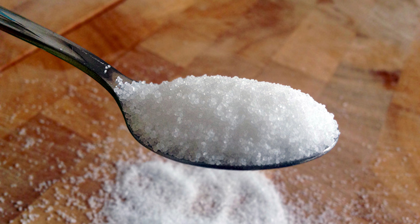
Sugar industry sought to sugarcoat causes of heart disease

Using records unearthed from library storage vaults, researchers recently revealed that the sugar industry paid nutrition experts from Harvard University to downplay studies linking sugar and heart disease. Although the incident happened in the 1960s, it appears to have helped redirect the scientific narrative for decades.

The documents — which include correspondence, symposium programs and annual reports — show that the Sugar Research Foundation (as it was named at the time) paid professors who wrote a two-part review in 1967 in the New England Journal of Medicine. That report was highly skeptical of the evidence linking sugar to cardiovascular problems but accepting of the role of fat. The now-deceased professors’ overall conclusion left “no doubt” that reducing the risk of heart disease was a matter of reducing saturated fat and cholesterol, according to researchers from the University of California, San Francisco, who published their report online September 12 in JAMA Internal Medicine.

“Why does it matter today? The sugar industry helped deflect the way the research was developing,” says study coauthor Cristin Kearns, a dentist at UCSF’s Institute for Health Policy Studies. The Harvard team’s scientific favoritism had a role in directing research and policy attention toward fat and cholesterol. And in fact, the first dietary guidelines published by the federal government in 1980 said there was no convincing evidence that sugar causes heart disease, stating “the major health hazard from too much sugar is tooth decay.”
Following the publication of the Harvard report, fat and cholesterol went on to hijack the scientific agenda for decades, and even led to a craze of low-fat foods that often added sugar. Kearns points out that it was only in 2015 that dietary guidelines finally made a strong statement to limit sugar. Researchers writing this year in Progress in Cardiovascular Diseases note that current studies estimate that diets high in added sugars carry a three times higher risk of death from cardiovascular disease. (For its part, the Sugar Association says in a statement on its website that “the last several decades of research have concluded that sugar does not have a unique role in heart disease.”)

The level at which the food industry continues to influence nutrition research is still a much-debated topic. The Sugar Association’s statement acknowledged the secret deal occurred, but pointed out that “when the studies in question were published, funding disclosures and transparency standards were not the norm they are today.” Journals now require all authors to list conflicts of interest, especially funding from a source has a vested interest in the outcome.

That doesn’t mean that trade groups and industry associations no longer have an influence on scientists, says Andy Bellatti, cofounder and strategic director of Dietitians for Professional Integrity, which has campaigned to push the Academy of Nutrition and Dietetics to sever its ties with industry, While a modern researcher could not take corporate money, even for speaking fees, without disclosure, the influences may be more subtle, he says. “We’re not talking about making up data, but perhaps influencing how a research question is framed.”

In a commentary published with the JAMA study, Marion Nestle, a nutrition researcher at New York University, wrote that industry influence has not disappeared. She cited recent New York Times investigations of Coca-Cola–sponsored research and Associated Press stories revealing that a candy trade group sponsored research attempting to show that children who eat sweets have a healthy body weight.

Bellatti says that researchers don’t necessarily want to be cozy with industry, but sometimes turn to commercial sources because non-biased research money is lacking. “The reason the food industry is able to do this is because there is such little public funding for nutrition and disease,” Bellatti says.
For that reason, the scientific community should not reject industry money wholesale, says John Sievenpiper, a physician and nutrition researcher at the University of Toronto. A study of his was once ridiculed on Nestle’s blog because the disclosures covered two full pages. He believes that any scientist who takes industry money should adhere to an even higher standard of openness, including releasing study protocols ahead of time so reviewers can make sure the research question was not changed midstream to favor a certain conclusion.

While many parallels have been made between the food and tobacco industries, Sievenpiper believes those comparisons miss the complicated nature of the human diet. Tobacco is always bad, never good. Sugar, fat, cholesterol and other components of diet are some of both, making research into their effects much more nuanced, he says. And unlike with tobacco, the solution can’t be to never eat them. He believes solutions won’t involve turning single nutrients like fat or sugar into villains, but promoting better overall patterns of eating, like the Mediterranean diet.

Kearns, who has spent the past 10 years looking into the sugar industry’s influence on science, isn’t finished yet. She says her curiosity first arose during a conference on gum disease and diabetes in 2007, when she noticed a lack of scientific discussion of sugar. She started out simply Googling industry influences. The trail eventually led her to scour library archives, until she came across dusty boxes of records from a closed sugar company in Colorado. “The first page I looked at in that archive had a confidential memo,” she says. “I knew I had something no one else had never talked about before.”

She doesn’t see the research going sour any time soon. “This was their 226th project in 1965,” she says. “There’s a lot more to the story.”

There’s a new way to stop an earthquake: put a volcano in its path

Editor’s note: Science has retracted the study described in this article. The May 3, 2019, issue of the journal notes that a panel of outside experts convened by Kyoto University in Japan concluded in March 2019 that the paper contained falsified data, manipulated images and instances of plagiarism, and that these were the responsibility of lead author Aiming Lin, a geophysicist at Kyoto University. In agreement with the investigation’s recommendation, the authors withdrew the report.

A titanic volcano stopped a mega-sized earthquake in its tracks.

In April, pent-up stress along the Futagawa-Hinagu Fault Zone in Japan began to unleash a magnitude 7.1 earthquake. The rupture traveled about 30 kilometers along the fault until it reached Mount Aso, one of Earth’s largest active volcanoes. That’s where the quake met its demise, geophysicist Aiming Lin of Kyoto University in Japan and colleagues report online October 20 in Science. The quake moved across the volcano’s caldronlike crater and abruptly stopped, the researchers found.

Geophysical evidence suggests that a region of rising magma lurks beneath the volcano. This magma chamber created upward pressure plus horizontal stresses that acted as an impassable roadblock for the seismic slip powering the quake, the researchers propose. This rare meetup, the researchers warn, may have undermined the structural integrity surrounding the magma chamber, increasing the likelihood of an eruption at Aso.

Young planets carve rings and spirals in the gas around their suns

Growing planets carve rings and spiral arms out of the gas and dust surrounding their young stars, researchers report in three papers to be published in Astronomy & Astrophysics. And dark streaks radiating away from the star in one of the planet nurseries appear to be shadows cast onto the disk by the clumps of planet-building material close to the star. This isn’t the first time that astronomers have spied rings around young stars, but the new images provide a peek at what goes into building diverse planetary systems.

The three stars — HD 97048, HD 135344B and RX J1615.3-3255 — are all youthful locals in our galaxy. They sit between 460 and 600 light-years away; the oldest is roughly a mere 8 million years old. All the stars have been studied before. But now three teams of researchers have used a new instrument at the Very Large Telescope in Chile to see extra-sharp details in the planet construction zone around each star.

The new instrument, named SPHERE, was designed to record images, spectra and polarimetry (the orientations of light waves) of young exoplanet families. Flexible mirrors within the instrument adapt to atmospheric turbulence above the telescope, and a tiny disk blocks light from the star, allowing faint details around the star to come into view.

There’s something cool about Arctic bird poop

Seabird poop helps the Arctic keep its cool, new research suggests.

The droppings release ammonia into the atmosphere, where it reacts with other chemicals in the air to form small airborne particles. Those particles form the heart of cloud droplets that reflect sunlight back into space, researchers propose November 15 in Nature Communications.

Even though the poop’s presence provides only modest cooling, understanding the effect could help scientists better predict how the region will fare under future climate change, says study coauthor Greg Wentworth. “The humor is not lost on me,” says Wentworth, an atmospheric chemist at Alberta Environment and Parks in Canada. “It’s a crucial connection, albeit somewhat comical.”
Arctic air temperatures are rising about twice as fast as temperatures in lower latitudes (SN: 12/26/15, p. 8), a shift that could threaten ecosystems and alter global weather patterns. Scientists still don’t fully understand Arctic climate, though.

Earlier this year, Wentworth and colleagues reported finding surprisingly abundant ammonia in Arctic air. They linked the chemical to the guano of the tens of millions of seabirds that flock to the frigid north each summer. Bacteria in the Arctic dine on the feces and release about 40,000 metric tons of ammonia annually. (The smell, Wentworth says, is awful.)

Once in the atmosphere, that ammonia reacts with sulfuric acid and water to form small particles that increase the number of cloud droplets, the researchers now propose. A cloud made up of a lot of smaller droplets will have more surface area and reflect more sunlight than a cloud made up of fewer but larger droplets.

This effect causes on average about 0.5 watts of summertime cooling per square meter in the Arctic, with more than a watt of cooling per square meter in some areas, the researchers estimate using a simulation of the Arctic’s atmospheric chemistry. For comparison, the natural greenhouse effect causes about 150 watts of warming per square meter worldwide. On top of that, carbon dioxide from human activities currently contributes about 1.6 watts per square meter of warming on average.

“Birds are in the equation now” when it comes to cloud formation, says Ken Carslaw, an atmospheric scientist at the University of Leeds in England. Understanding how climate change and human activities in the Arctic impact seabirds could be important to forecasting future temperature changes in the region, he says.

Bacteria help carnivorous plants drown their prey

Bacteria may be a meat-eating plant’s best friends thanks to their power to reduce the surface tension of water.

The carnivorous pitcher plant Darlingtonia californica releases water into the tall vases of its leaves, creating deathtraps where insect prey drown. Water in a pitcher leaf starts clear. But after about a week, thanks to bacteria, it turns “murky brown to a dark red and smells horrible,” says David Armitage of the University of Notre Dame in Indiana. Now, he’s found that those bacteria can help plants keep insects trapped. Microbial residents reduce the surface tension of water enough for ants and other small insects to slip immediately into the pool instead of perching lightly on the surface, he reports November 23 in Biology Letters.

Armitage seeded tubes of clean water with fluid from the trap pools of pitcher plants and added dead crickets to feed the microbes. After sitting for a month, the mess had about the same surface tension properties as natural pitcher plant pools. Then, he created a series of increasingly dilute samples of pool soup and dropped harvester ants into each one. He found that the ants sank immediately in all but the bacteria-free water sample.

Bacterial populations in a pitcher leaf are akin to those in a mammal gut or bovine rumen, Armitage’s preliminary analysis finds. The microbes can help digest the prey as well as catch it, he says.

Cell biologists learn how Zika kills brain cells, devise schemes to stop it

SAN FRANCISCO — Cell biologists are learning more about how the Zika virus disrupts brain cells to cause the birth defect microcephaly, in which a baby’s brain and head are smaller than usual. Meantime, several strategies to combat the virus show preliminary promise, researchers reported at the American Society for Cell Biology’s annual meeting. Among the findings:

Brain cell die-off
Zika causes fetal brain cells neighboring an infected cell to commit suicide, David Doobin of Columbia University Medical Center reported December 6. In work with mice and rats, Doobin and colleagues found suggestions that the cells’ death might be the body’s attempt to limit spread of the virus.

The researchers applied techniques they had previously used to investigate a genetic cause of microcephaly to narrow when in pregnancy the virus is most likely to cause the brain to shrink. Timing of the virus’s effect varied by strain. For one from Puerto Rico, brain cell die-off happened in mice only in the first two trimesters. But a strain from Honduras could kill developing brain cells later into pregnancy. Microcephaly can lead to seizures, mental impairment, delays in speech and movement and other problems.

Enzyme stopper
Disrupting a Zika enzyme could help stop the virus. The enzyme, NS3, causes problems when it gloms on to centrioles, a pair of structures inside cells needed to divvy up chromosomes when cells divide, Andrew Kodani, a cell biologist at Boston Children’s Hospital reported December 6.

Zika, dengue and other related viruses, known as flaviviruses, all use a version of NS3 to chop joined proteins apart so they can do their jobs. (Before chopping, Zika’s 10 proteins are made as one long protein.) But once NS3 finishes slicing virus proteins, the enzyme moves to the centrioles, where it can mess with their assembly, Kodani and colleagues found. Something similar happens in some genetic forms of microcephaly.

A chemical called an anthracene can help fend off dengue, so Kodani and colleagues tested anthracene on Zika as well. Small amounts of the chemical can prevent NS3 from tinkering with the centrioles, the researchers found. So far the work has only been done in lab dishes.
Protein face-off
Another virulent virus could disable Zika. Work with cells grown in lab dishes suggests a bit of protein, or peptide, from the hepatitis C virus, could muck up Zika’s proteins.

The peptide interferes with HSP70, a protein that helps assemble complexes of other proteins, including ones involved in protein production. That peptide and other compounds were already known to inhibit hepatitis C replication, UCLA virologist Ronik Khachatoorian and colleagues had previously discovered. The hepatitis C virus peptide stops Zika virus proteins from being made and hampers assembly of the virus, Khachatoorian reported December 5.

The peptide has not been tested in animals yet.

Cells snack on nanowires

Human cells can snack on silicon.

Cells grown in the lab devour nano-sized wires of silicon through an engulfing process known as phagocytosis, scientists report December 16 in Science Advances.

Silicon-infused cells could merge electronics with biology, says John Zimmerman, a biophysicist now at Harvard University. “It’s still very early days,” he adds, but “the idea is to get traditional electronic devices working inside of cells.” Such hybrid devices could one day help control cellular behavior, or even replace electronics used for deep brain stimulation, he says.
Scientists have been trying to load electronic parts inside cells for years. One way is to zap holes in cells with electricity, which lets big stuff, like silicon nanowires linked to bulky materials, slip in. Zimmerman, then at the University of Chicago, and colleagues were looking for a simpler technique, something that would let tiny nanowires in easily and could potentially allow them to travel through a person’s bloodstream — like a drug.
Zimmerman’s team had previously shown that cells could take in silicon nanowires, but no one knew how it worked. So he added the nanowires to different kinds of cells — including human umbilical vein cells, rat nerve cells and mouse immune cells — in laboratory dishes. Under a microscope, Zimmerman says, “you can see the cell grab the wire, wrap a membrane around it, and pull it inside — kind of like a lasso.” Then, the wire travels on molecular tracks through the cell’s interior to settle around the nucleus.
Molecular tests suggested that nanowires entered via phagocytosis, a process by which some cells take in bacteria and cellular junk. During phagocytosis, a cell’s membrane encapsulates the junk, forming a pouch that carries the cargo to a recycling center inside the cell.

Not all cell types swallowed the wires, though. Knowing which types do, and how the wires get inside is important, Zimmerman says, because it could help in predicting where they would end up in the body.
But there’s still a long way to go from nanowire-loaded cells to working electronic devices, says Mark Reed, a physicist at Yale University. “This is the big question,” he says — because the nanowires aren’t actually hooked up to anything yet.

Meat-eating pitcher plants raise deathtraps to an art

Tricking some bug into drowning takes finesse, especially for a hungry meat eater with no brain, eyes or moving parts. Yet California pitcher plants are very good at it.

Growing where deposits of the mineral serpentine would kill most other plants, Darlingtonia californica survives in low-nutrient soil by being “very meat dependent,” says David Armitage of the University of Notre Dame in Indiana. Leaves he has tested get up to 95 percent of their nitrogen from wasps, beetles, ants or other insects that become trapped inside the snake-curved hollow leaves.
The leaves don’t collect rainwater because a green dome covers the top. Instead, they suck moisture up through the roots and (somehow) release it into the hollow trap. “People have been doing weird experiments where they feed [a plant] meat and milk and other things to try to trigger it to release water,” Armitage says. Experiments tempting the green carnivore with cheese, beef broth, egg whites and so on suggest there’s some sort of chemical cue.

However the water enters the leaf pool, it starts out clear. As insects drown, the liquid darkens to a murky brown or red and “smells just horrible,” he says. The soupiness comes from bacteria, which help doom prey by lowering the surface tension of the drowning pool, Armitage reports in the November Biology Letters. Ants or other small insects sink below the surface immediately instead of floating at the top.
But first, pitchers lure victims to the pool by repurposing an old plant ploy: free nectar. It’s “highly nitrogen-rich and full of sugars, so it’s delicious — I’ve tasted it,” Armitage says. Pitcher plants sprout blooms, but the trap nectar doesn’t come from the drooping flowers. A roll of tissue near the pitcher mouth oozes the treat.

That nectar-heavy roll curves onto what’s called the fishtail appendage. Mature plants
(2 years or older) grow this forked tissue like a moustache at the pitcher mouth. Biologists for more than a century have presumed that this big, red-veined, lickable prong worked as an insect lure. Armitage, however, tested the idea and says it may be wrong.
Clipping fishtails off individual leaves, or even off all the leaves in a small patch, did nothing to shrink the catch compared with fully mustachioed leaves, he reported in the American Journal of Botany in April 2016. The only thing fishtails lure, for the time being at least, are puzzled botanists.

Coastal waters were an oxygen oasis 2.3 billion years ago

Earth was momentarily ripe for the evolution of animals hundreds of millions of years before they first appeared, researchers propose.

Chemical clues in ancient rocks suggest that 2.32 billion to 2.1 billion years ago, shallow coastal waters held enough oxygen to support oxygen-hungry life-forms including some animals, researchers report the week of January 16 in the Proceedings of the National Academy of Sciences. But the first animal fossils, sponges, don’t appear until around 650 million years ago, following a period of scant oxygen known as the boring billion (SN: 11/14/15, p. 18).
“As far as environmental conditions were concerned, things were favorable for this evolutionary step to happen,” says study coauthor Andrey Bekker, a sedimentary geologist at the University of California, Riverside. Something else must have stalled the rise of animals, he says.

Microbes began flooding Earth with oxygen around 2.3 billion years ago during the Great Oxidation Event. This breath of oxygen enabled the eventual emergence of complex, oxygen-dependent life-forms called eukaryotes, an evolutionary line that would later include animals and plants. Scientists have proposed that the Great Oxidation Event wasn’t a smooth rise, but contained an “overshoot” during which oxygen concentrations momentarily peaked before dropping to a lower, stable level during the boring billion. Whether that overshoot was enough to support animals was unclear, though.

Bekker and colleagues tackled this question using a relatively new way to measure ancient oxygen. Rock weathering can wash the element selenium into the oceans. In oxygen-free waters, all of the selenium settles onto the seafloor. But in water with at least some oxygen, only a fraction of the selenium is deposited. And the selenium that is laid down is disproportionately that of a lighter isotope of the element, leaving atoms of a heavier isotope to be deposited elsewhere.
If ancient coasts contained relatively abundant oxygen, the researchers expected to find more light selenium close to shore and more heavy selenium in deeper, oxygen-deprived waters. Analyzing shales formed under deep waters around the world, the researchers found just such an isotope segregation. These shales had an abundance of the heavier selenium, leading researchers to infer that the lighter version of the element was concentrated closer to shore.

Oxygen concentrations in coastal waters were at least nearly one percent of present-day levels and “were flirting with the limits of what complex life can survive,” proposes study coauthor Michael Kipp, a geochemist at the University of Washington in Seattle. While the environment was probably suitable for eukaryotes, life hadn’t evolved enough by this point to take advantage of the situation, Kipp proposes. The appearance of eukaryotes in the fossil record would take hundreds of millions of years of more evolution (SN Online: 5/18/16), he says, and the first animals even longer.
Tracking selenium is such a new technique, though, that “interpretations could change as we better understand how it works,” says Philip Pogge von Strandmann, a geochemist at University College London. Currently the method is “tricky,” he says, especially for precisely estimating oxygen concentrations.

Construction of tiny, fluid-filled devices inspired by Legos

Legos have provided the inspiration for small, fluid-ferrying devices that can be built up brick-by-brick.

Tools for manipulating tiny amounts of liquid, known as microfluidic devices, can be used to perform blood tests, detect contaminants in water or simulate biological features like human blood vessels. The devices are easily portable, about the size of a quarter and require only small samples of liquid.

But fabricating such devices is not easy. Each new application requires a different configuration of twisty little passages, demanding a brand new design that must be molded or 3-D printed.

Scientists from the University of California, Irvine created Lego-style blocks out of a polymer called PDMS. Their bricks contained minuscule channels, half a millimeter wide, that allowed liquid to flow from brick to brick with no leaks. New devices could be created quickly by rearranging standard blocks into various configurations, the scientists report January 24 in the Journal of Micromechanics and Microengineering.