Fossils hint at India’s crucial role in primate evolution

Remarkably preserved bones of rat-sized creatures excavated in an Indian coal mine may come from close relatives of the first primatelike animals, researchers say.

A set of 25 arm, leg, ankle and foot fossils, dating to roughly 54.5 million years ago, raises India’s profile as a possible hotbed of early primate evolution, say evolutionary biologist Rachel Dunn of Des Moines University in Iowa and her colleagues. Bones from Vastan coal mine in Gujarat, India’s westernmost state, indicate that these tiny tree-dwellers resembled the first primates from as early as 65 million years ago, the scientists report in the October Journal of Human Evolution.
These discoveries add to previously reported jaws, teeth and limb bones of four ancient primate species found in the same mine. “The Vastan primates probably approximate a common primate ancestor better than any fossils found previously,” says paleontologist and study coauthor Kenneth Rose of Johns Hopkins University School of Medicine.
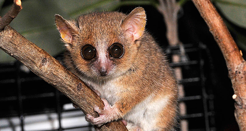
The Vastan animals were about the size of living gray mouse lemurs and dwarf lemurs, weighing roughly 150 to 300 grams (roughly half a pound), the investigators estimate. Dunn’s group has posted 3-D scans of the fossils to Morphosource.org ( SN: 3/19/16, p. 28 ) so other researchers can download and study the material.
Most Vastan individuals possessed a basic climbing ability unlike the more specialized builds of members of the two ancient primate groups that gave rise to present-day primates, the researchers say. One of those groups, omomyids, consisted of relatives of tarsiers, monkeys and apes. The other group, adapoids, included relatives of lemurs, lorises and bushbabies. The Indian primates were tree-dwellers but could not leap from branch to branch like lemurs or ascend trees with the slow-but-sure grips of lorises, the new report concludes.

Vastan primates probably descended from a common ancestor of omomyids and adapoids, the researchers propose. India was a drifting landmass headed north toward a collision with mainland Asia when the Vastan primates were alive. Isolated on a huge chunk of land, the Indian primates evolved relatively slowly, retaining a great number of ancestral skeletal traits, Rose suspects.

“It’s possible that India played an important role in primate evolution,” says evolutionary anthropologist Doug Boyer of Duke University. A team led by Boyer reported in 2010 that a roughly 65-million-year-old fossil found in southern India might be a close relative of the common ancestor of primates, tree shrews and flying lemurs (which glide rather than fly and are not true lemurs).

One possibility is that primates and their close relatives evolved in isolation on the island continent of India between around 65 million and 55 million years ago, Boyer suggests. Primates then spread around the world once India joined Asia by about 50 million years ago.

That’s a controversial idea. An increasing number of scientists suspect primates originated in Asia. Chinese primate fossils dating to 56 million to 55 million years ago are slightly older than the Vastan primates (SN: 6/29/13, p. 14; SN: 1/3/04, p. 4). The Chinese finds show signs of having been omomyids.

And in at least one respect, Boyer says, some of the new Vastan fossils may be more specialized than their discoverers claim. Vastan ankle bones, for instance, look enough like those of modern lemurs to raise doubts that the Indian primates were direct descendants of primate precursors, he holds.

Dunn, however, regards the overall anatomy of the Vastan fossils as “the most direct evidence we have” that ancestors of early primates lacked lemurs’ leaping abilities, contrary to what some researchers have argued.

How one scientist’s gut microbes changed over a year

Where you live and what you eat can rapidly affect the types of friendly bacteria inhabiting your body. To see how the microbes that inhabit the mouth and intestines change over time, Duke University computational biologist Lawrence David zealously chronicled his microbiome for an entire year. (For more on David and this experiment, see “Lawrence David’s gut check gets personal.”)

A stream plot (below, top graph) shows the ebb and flow of phyla of bacteria in his gut over time. The thickness of each stream indicates a bacterial group’s relative abundance in daily fecal samples.
David peered closer at the data in a horizon plot (above, bottom graph; colored squares at left indicate the phylum of the bacteria represented in each row). He first determined each type of bacteria’s normal abundance in his gut, then calculated how much they differed from the median abundance. Warmer colors (red, orange, yellow) indicate that bacteria in that group increased in abundance, and cooler colors (blue, green) indicate a decrease in abundance. Living abroad from day 71 to day 122 had a dramatic — but short-lived — effect on David’s microbiome.

Sugar industry sought to sugarcoat causes of heart disease

Using records unearthed from library storage vaults, researchers recently revealed that the sugar industry paid nutrition experts from Harvard University to downplay studies linking sugar and heart disease. Although the incident happened in the 1960s, it appears to have helped redirect the scientific narrative for decades.

The documents — which include correspondence, symposium programs and annual reports — show that the Sugar Research Foundation (as it was named at the time) paid professors who wrote a two-part review in 1967 in the New England Journal of Medicine. That report was highly skeptical of the evidence linking sugar to cardiovascular problems but accepting of the role of fat. The now-deceased professors’ overall conclusion left “no doubt” that reducing the risk of heart disease was a matter of reducing saturated fat and cholesterol, according to researchers from the University of California, San Francisco, who published their report online September 12 in JAMA Internal Medicine.

“Why does it matter today? The sugar industry helped deflect the way the research was developing,” says study coauthor Cristin Kearns, a dentist at UCSF’s Institute for Health Policy Studies. The Harvard team’s scientific favoritism had a role in directing research and policy attention toward fat and cholesterol. And in fact, the first dietary guidelines published by the federal government in 1980 said there was no convincing evidence that sugar causes heart disease, stating “the major health hazard from too much sugar is tooth decay.”
Following the publication of the Harvard report, fat and cholesterol went on to hijack the scientific agenda for decades, and even led to a craze of low-fat foods that often added sugar. Kearns points out that it was only in 2015 that dietary guidelines finally made a strong statement to limit sugar. Researchers writing this year in Progress in Cardiovascular Diseases note that current studies estimate that diets high in added sugars carry a three times higher risk of death from cardiovascular disease. (For its part, the Sugar Association says in a statement on its website that “the last several decades of research have concluded that sugar does not have a unique role in heart disease.”)

The level at which the food industry continues to influence nutrition research is still a much-debated topic. The Sugar Association’s statement acknowledged the secret deal occurred, but pointed out that “when the studies in question were published, funding disclosures and transparency standards were not the norm they are today.” Journals now require all authors to list conflicts of interest, especially funding from a source has a vested interest in the outcome.

That doesn’t mean that trade groups and industry associations no longer have an influence on scientists, says Andy Bellatti, cofounder and strategic director of Dietitians for Professional Integrity, which has campaigned to push the Academy of Nutrition and Dietetics to sever its ties with industry, While a modern researcher could not take corporate money, even for speaking fees, without disclosure, the influences may be more subtle, he says. “We’re not talking about making up data, but perhaps influencing how a research question is framed.”

In a commentary published with the JAMA study, Marion Nestle, a nutrition researcher at New York University, wrote that industry influence has not disappeared. She cited recent New York Times investigations of Coca-Cola–sponsored research and Associated Press stories revealing that a candy trade group sponsored research attempting to show that children who eat sweets have a healthy body weight.

Bellatti says that researchers don’t necessarily want to be cozy with industry, but sometimes turn to commercial sources because non-biased research money is lacking. “The reason the food industry is able to do this is because there is such little public funding for nutrition and disease,” Bellatti says.
For that reason, the scientific community should not reject industry money wholesale, says John Sievenpiper, a physician and nutrition researcher at the University of Toronto. A study of his was once ridiculed on Nestle’s blog because the disclosures covered two full pages. He believes that any scientist who takes industry money should adhere to an even higher standard of openness, including releasing study protocols ahead of time so reviewers can make sure the research question was not changed midstream to favor a certain conclusion.

While many parallels have been made between the food and tobacco industries, Sievenpiper believes those comparisons miss the complicated nature of the human diet. Tobacco is always bad, never good. Sugar, fat, cholesterol and other components of diet are some of both, making research into their effects much more nuanced, he says. And unlike with tobacco, the solution can’t be to never eat them. He believes solutions won’t involve turning single nutrients like fat or sugar into villains, but promoting better overall patterns of eating, like the Mediterranean diet.

Kearns, who has spent the past 10 years looking into the sugar industry’s influence on science, isn’t finished yet. She says her curiosity first arose during a conference on gum disease and diabetes in 2007, when she noticed a lack of scientific discussion of sugar. She started out simply Googling industry influences. The trail eventually led her to scour library archives, until she came across dusty boxes of records from a closed sugar company in Colorado. “The first page I looked at in that archive had a confidential memo,” she says. “I knew I had something no one else had never talked about before.”

She doesn’t see the research going sour any time soon. “This was their 226th project in 1965,” she says. “There’s a lot more to the story.”

CT scans show first X-rayed mummy in new light

X-rays were the iPhone 7 of the 1890s. Months after X-rays were discovered in late 1895, German physicist Walter Koenig put the latest in tech gadgetry to the test by scanning 14 objects, including the mummified remains of an ancient Egyptian child. Koenig’s image of the child’s knees represented the first radiographic investigation of a mummy.

At the time, details on the mummy itself were scant. Originally collected by explorer-naturalist Eduard Rueppell in 1817, the specimen lacked any sort of decoration that might link it to a particular dynasty or time period. Koenig’s X-ray image of the mummy served less to fill in any of those blanks and more to demonstrate the technology’s potential. Since then, radiographic images have revealed hidden artifacts, elucidated embalming techniques and even pinpointed health issues and diseases in mummies.
Now, biological anthropologist and Egyptologist Stephanie Zesch of the Reiss Engelhorn Museum in Mannheim, Germany, and colleagues have examined the mummy with modern imaging techniques. CT scans show that the child was a boy. His teeth suggest that he was 4 to 5 years old when he died. Radiocarbon dating places him in the Ptolemaic period, between 378 and 235 B.C., the researchers report online July 22 in the European Journal of Radiology Open.
The team also diagnosed a slew of health conditions: a common chest wall deformity called pectus excavatum, or sunken chest; bone density marks called Harris lines in his leg bones that indicate physiological stress; and an enlarged liver. The team attributes the distended liver to a parasitic infection like schistosomiasis, which is common in Egypt and sometimes lethal. Without any obvious signs of trauma, however, “it’s impossible to determine cause of death,” Zesch says.

Even with the all-seeing power of today’s CT scans, the culprit behind the boy’s demise remains under wraps.